flow cytometry results interpretation
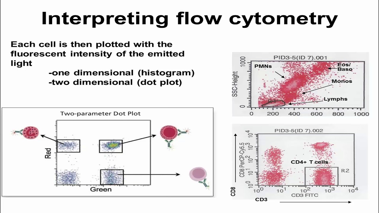
1 identification of cells from different lineages and. The iQue 3 combines a patented sampling method which allows for the fastest sample acquisition in the industry.
Introduction To Flow Cytometric Analysis Flow Cytometry
The flow cytometry data that forms the basis of the conclusions should be presented clearly and concisely.

. To get you from samples to actionable results in record time. Reference ranges for blood tests are 32 to 36 gdL 320 to 360gL or between 481 and 558 mmolL. It is calculated by dividing the hemoglobin by the hematocrit.
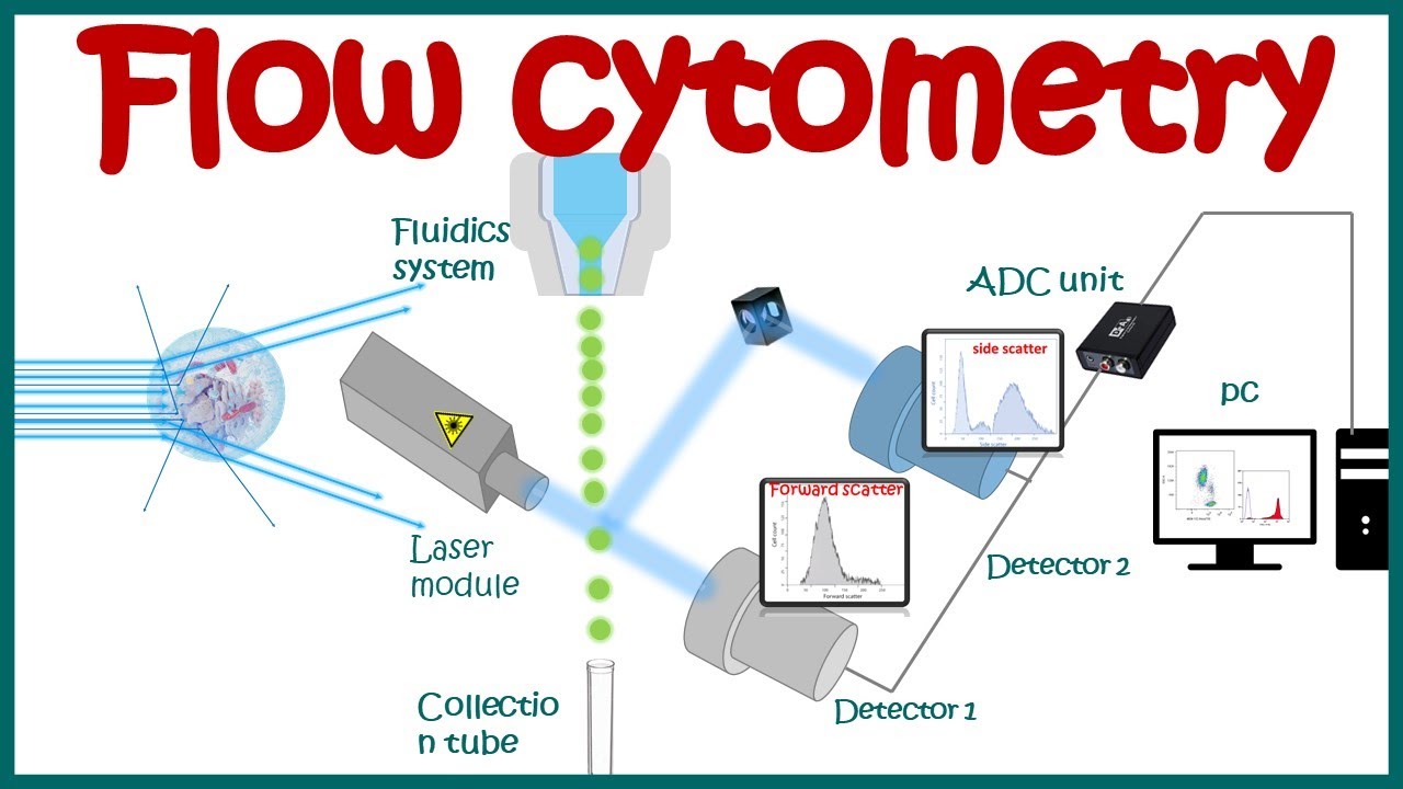
First described about 70 years ago it is elegantly simple in design yet it is widely complex in its applications and interpretations and it is prone to false-positive and false-negative results. This chapter discusses these components. MitoSOX-based flow cytometry is a quick simple and inexpensive method for detecting cellular ROS.
Here are the. Release of Exam Score to Program Officials. It has the ability to handle 96 384 well plates and.
From testing and interpretation of results to consultation services we provide our clients with information to prevent or treat veterinary issues for a wide variety of animal species. Since Medicare does not pay for duplicate testing do not report flow cytometry on multiple specimens on the same date of service unless the morphology or other clinical factors suggest differing results on the. Remember to acknowledge the valuable services provided by the Cell Analysis Facility in your research papers and publications.
Peer-reviewed articles cover topics in oncology trauma gastrointestinal vascular and transplantation surgeryThe journal also. Examination results cannot be released by telephone or fax to anyone. For 66 years Surgery has published practical authoritative information about procedures clinical advances and major trends shaping general surgeryEach issue features original scientific contributions and clinical reports.
The most advanced flow cytometry platform with a focus on speed from setup to the acquisition and analysis. These results suggest that RA disease activity measured weekly with a validated patient-reported outcome is stable around the time of an AddDose of COVID-19 vaccine. If the probe is used at an optimal concentration MitoSOX-based flow cytometry can reliably detect the relative differences in mitochondrial ROS formation in cells.
Measurable residual disease was determined by central assessment with flow cytometry with the use of a leukemia-associated immunophenotype LAIPbased. Here we will walk you through specific flow cytometry assays with examples that will enable. Flow cytometric immunophenotyping evaluates individual cells in suspension for the presence and absence of specific antigens phenotype.
The number of participants providing blood for flow cytometry analyses was low particularly in subgroups by holding versus continuing DMARDs and results should be interpreted cautiously. Send your request by mail to. Quickly set up runs and monitor amplification traces in real time on the integrated LCD touch screen or use the included CFX Maestro Software to easily and intuitively design your experiment and analyze results from a connected computer.
Flow cytometry interpretation should be reported using CPT codes 88187-88189. The mean corpuscular hemoglobin concentration MCHC is a measure of the concentration of hemoglobin in a given volume of packed red blood cell. Careful experimental set-up and interpretation of results will allow you to make the most of your experiment.
Two fully accredited locations to serve you. Flow cytometric leukemia and lymphoma analysis may aid in identifying the tumor lineage for diagnostic and prognostic purposes. For information on South Africas response to COVID-19 please visit the COVID-19 Corona Virus South African Resource Portal.
In addition to providing access to the flow cytometers and cell sorting services we provide technical support to assist in experimental design data analysis and interpretation as well as development of novel flow cytometric based techniques. Only one code should be reported for all flow cytometry performed on a specimen. With up to five-target detection unsurpassed thermal cycler performance unrivaled stand-alone functionality and powerful yet easy-to-use.
When preparing figures for publication the scientific question and hypothesis that forms the basis of the paper must be central and all the figures must be in support of that. Our comprehensive reagent portfolio includes clinical diagnostic testing kits kits for innovative new approaches to clinical research and single-color reagents for research clinical research or lab-developed. Components of the laboratory exam include complete blood count with differential comprehensive metabolic panel inflammatory markers autoantibodies and flow cytometry.
Bosters mission is to support research in areas such as immunology neuroscience cancer and more by providing the high-quality ELISA kits needed to get better results. In most cases the lineage can be identified as T-cell B-cell or myeloid and a diagnosis or. The direct antiglobulin test DAT.
It is thus a mass or molar concentrationStill many instances measure. The results can confirm a diagnosis estimate disease severity aid in assessing prognosis and are useful to follow disease activity. If you completed a NAACLS ABHES or CAAHEP accredited program your examination scores will be released to the officials of your program unless you instruct the ASCP Board of Certification otherwise.
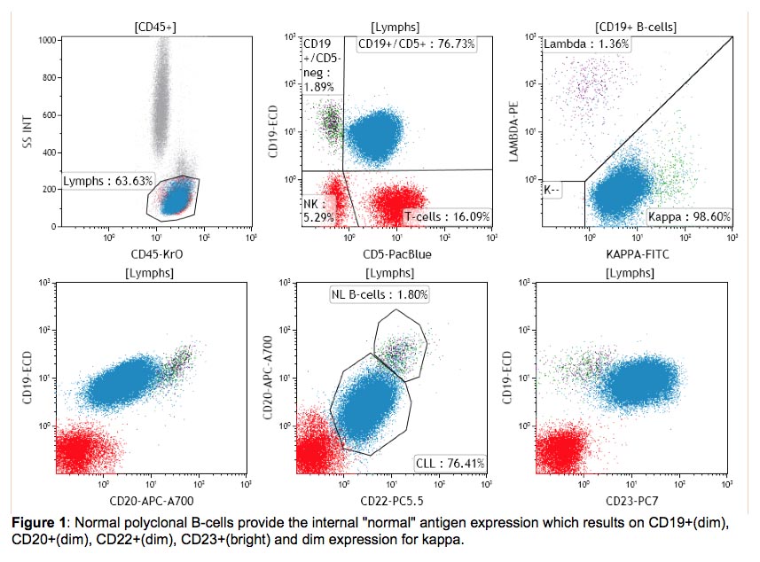
Both the Athens and Tifton Diagnostic Laboratories are fully accredited by the American Association of Veterinary Laboratory Diagnosticians. After review of the clinical history and morphology a panel of markers is selected for each case by a board-certified hematopathologist. While it provides pretty pictures and colorful layouts the meat of the data are the numbers.
Sometimes referred to as the Coombs test continues to be one of the most widely used assays in laboratory medicine. Flow Cytometry Resource Facility. This research was supported by the Cell Analysis Facility Flow Cytometry Shared Resource Lab in the Department of Immunology at the University of Washington CAF COVID-19 Policies.
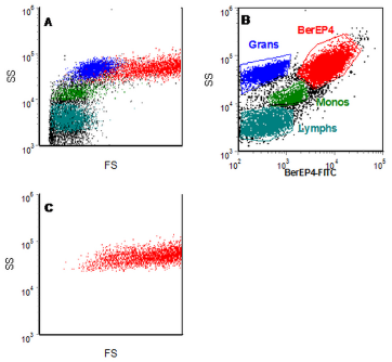
BD Biosciences flow cytometry reagents truly reflect our scientific leadership in flow cytometry innovation and our 45 years of dedication to providing high-quality products. Flow cytometry allows the study of all aspects of apoptosis from induction via surface receptors to late stages where DNA fragmentation occurs. In the assessment for hematologic malignancies several steps are taken in the application and interpretation of this immunophenotypic information.
Our 2300 ELISA kits are validated in multiple sample matrices from serum and saliva to urine and feces ensuring wide application ranges for you to select from. It is suggested that 1 μM instead of 5 μM of MitoSOX be used as the working.
Data Analysis What Does A Histogram For Flow Cytometry Tell Me Unsolved Mysteries Of Human Health Oregon State University
Flow Cytometry Planning Assignment

Basics Of Flow Cytometry Part I Gating And Data Analysis Youtube

Basic Or Advanced Flow Cytometry Webinar Training

Overview Of High Dimensional Flow Cytometry Data Analysis A Fcs Download Scientific Diagram

Flow Cytometry Basics Flow Cytometry Miltenyi Biotec Technologies Macs Handbook Resources Miltenyi Biotec Usa
Flow Cytometry Basic Principles What The Use Of Flow Cytometry Cell Sorting By Facs Youtube

Show Dot Blot Analysis Of Flow Cytometry Data Of Cd4 Cd8 Of Two Cases Download Scientific Diagram

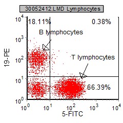
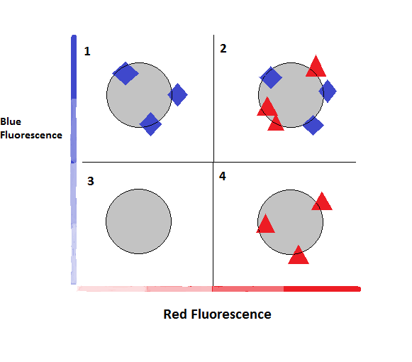
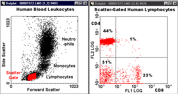
Chapter 4 Data Analysis Flow Cytometry A Basic Introduction
Chapter 4 Data Analysis Flow Cytometry A Basic Introduction

6 Areas Of Consideration For Flow Cytometry Cell Cycle Analysis Cheeky Scientist
Introduction To Flow Cytometric Analysis Flow Cytometry
International Clinical Cytometry Society

Flow Cytometry Tutorial Flow Cytometry Data Analysis Flow Cytometry Gating Youtube
Flow Cytometry Verbsky Youtube

How To Analyze Flow Cytometry Data
A D Flow Cytometry Interpretation The Neoplastic Cells Display The Download Scientific Diagram

2 An Example Of Flow Cytometry Data Analysis Facs Purification The Download Scientific Diagram
Blog Flow Cytometry Data Analysis I What Different Plots Can Tell You